
How a Growth Scan in Pregnancy Monitors Baby Development
Medically reviewed by Mr Amer Raza MBBS MRCOG, Consultant Gynaecologist, Ovara Health (GMC 5205372) | Last reviewed: 4 July 2026
Key Facts: A growth scan in pregnancy is usually performed in the third trimester to check the baby’s estimated weight, growth pattern, amniotic fluid and placental function. It is recommended if growth appears different from expected, movements reduce, or there are risk factors such as diabetes or high blood pressure. The scan is safe, non-invasive and usually takes 20 to 30 minutes.
A growth scan in pregnancy is an ultrasound performed in the second half of pregnancy to assess the baby’s size, development and overall wellbeing. It measures key fetal parameters, checks amniotic fluid levels and evaluates placental function, helping to confirm the baby is growing appropriately and receiving adequate support inside the womb.
Unlike the 12-week scan and anomaly scan, which confirm dating and check the baby’s structure, a growth scan focuses on monitoring how the baby is growing over time. It is particularly important when there are risk factors such as high blood pressure, gestational diabetes, reduced fetal movements or previous pregnancy complications.
Quick Answer: A growth scan checks how well your baby is growing by measuring the head, abdomen and thigh bone to estimate weight, alongside amniotic fluid and placental blood flow. It is most often done from 28 weeks onwards, more frequently if there are concerns, and takes around 20 to 30 minutes.
When is a growth scan needed?
A growth scan is usually arranged when there are clinical concerns about the baby’s growth or the mother’s health. It is commonly recommended in the third trimester, particularly if routine measurements suggest the baby may be smaller or larger than expected for their stage of pregnancy.
A scan may be advised if there is reduced fetal movement, high blood pressure, gestational diabetes, a previous baby with growth restriction, a twin pregnancy, or concerns raised during a routine antenatal appointment. It may also be arranged if the size of the womb measures differently from expected on examination. The aim is to assess fetal size, amniotic fluid and placental function to guide further monitoring or delivery planning.
What does a growth scan check?
A growth scan involves measuring the baby’s head circumference, abdominal circumference and femur (thigh bone) length. These measurements are used to estimate fetal weight and are plotted on a growth chart against gestational age. The scan also assesses:
- Placental position and appearance
- Amniotic fluid volume
- Umbilical cord blood flow (Doppler)
- Fetal movements and breathing activity
The findings are documented in a growth scan report and interpreted in relation to gestational age and your individual clinical picture, not against a single fixed number.
What happens at each stage of pregnancy?
Understanding growth scan weeks helps explain why repeated scans may be recommended as pregnancy progresses.
24 to 28 weeks
A scan at this stage may be advised in selected cases where earlier monitoring is required, for example with a twin pregnancy or a relevant medical history. The focus is on establishing a growth baseline and confirming that fetal size matches gestational age. Between 24 and 28 weeks, doctors assess early signs of growth restriction, placental function and amniotic fluid levels. By 28 weeks, fetal weight estimation becomes more reliable, and this stage helps detect babies who are measuring smaller or larger than expected.
28 to 32 weeks
A scan is often scheduled in this window if concerns exist, with the baby’s weight gain pattern carefully monitored. Clinicians look for consistent growth along the centile chart, signs of placental insufficiency, and adequate blood flow on Doppler studies. If results show a deviation from the expected pattern, more frequent monitoring may be recommended.
32 to 36 weeks
This is a crucial phase of fetal development. A scan here provides important information about estimated birth weight and readiness for delivery. Between 32 and 36 weeks, the scan evaluates fetal growth progression, amniotic fluid stability, placental maturity and fetal position. Abnormal findings at this stage may start to influence delivery planning.
36 to 38 weeks
A scan may be advised in this window if there are concerns about reduced movements, diabetes or blood pressure disorders. The focus shifts to ensuring continued adequate growth, monitoring placental blood flow and confirming the baby’s position. This stage helps determine whether labour can proceed naturally or whether earlier intervention may be required.
38 to 40 weeks
A scan is sometimes performed in this window if pregnancy continues close to or beyond the due date, to help assess whether the placenta is still functioning efficiently. Doctors monitor for signs of placental ageing and overall fetal wellbeing. If fluid levels decrease or blood flow changes are detected, induction of labour may be considered.
How accurate is a growth scan?
Growth scans are generally reliable when performed by experienced professionals, but it is important to understand that estimated fetal weight is calculated from measurements and carries a recognised margin of error, typically around 10 to 15%. The real value of a growth scan lies in tracking growth trends over time, across more than one scan, rather than relying on a single measurement in isolation.
What if my baby measures small or large?
A single measurement slightly above or below average is not automatically a cause for concern; every baby has a different growth potential. Doctors look at the pattern of growth over time and your individual health, not one number in isolation.
If a baby measures consistently small for their gestational age, this may be described as fetal growth restriction, and can be linked to placental function, maternal blood pressure or other factors. It may lead to closer monitoring, additional Doppler scans, or a discussion about the right timing for delivery. If a baby measures large for their gestational age, this can sometimes be linked to gestational diabetes and may prompt a discussion about blood sugar screening and delivery planning. In both cases, the scan is there to guide safe decisions, not to alarm you, and most babies flagged on a single scan go on to be perfectly healthy.
Noticed your baby moving less than usual? Do not wait for your next scheduled scan. Call us on 0207 751 4488 or get in touch for prompt assessment. Reduced fetal movement should always be checked the same day.
Book a growth scan in Chelsea →
What happens during a growth scan appointment?
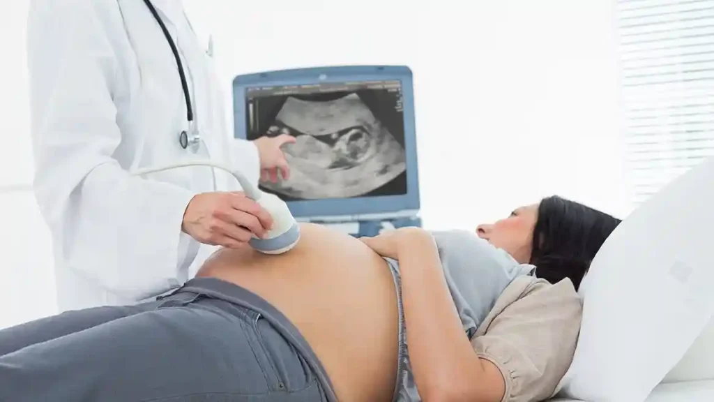
A growth scan is similar to other pregnancy ultrasounds. Gel is applied to the abdomen and a probe is moved across the skin to capture images; the examination is non-invasive and safe for both mother and baby. The sonographer takes measurements, checks fluid levels, and may assess blood flow using Doppler if indicated. The scan usually lasts between 20 and 30 minutes, though this can vary depending on fetal position and whether Doppler studies are needed. Findings are recorded in a growth scan report and discussed with you at the appointment.
Growth scan at Ovara Health, Chelsea
A growth scan plays an important role in monitoring fetal development during the later stages of pregnancy. By assessing fetal size, amniotic fluid and placental function at the right growth scan weeks, our Consultants can detect concerns early and guide safe management. While growth scan accuracy has natural limitations, it remains a valuable part of modern antenatal care. No GP referral is needed to book at Ovara Health, and same-week appointments are usually available.
Frequently asked questions
What is a growth scan in pregnancy?
A growth scan is an ultrasound, usually performed in the third trimester, that measures the baby’s head, abdomen and thigh bone to estimate weight and growth, alongside amniotic fluid levels and placental function.
When is a growth scan usually done?
Growth scans are usually performed from around 28 weeks onwards, with the exact timing and frequency depending on your individual risk factors and clinical findings.
Why would I need a growth scan?
Common reasons include reduced fetal movement, high blood pressure, gestational diabetes, a twin pregnancy, previous growth restriction, or a difference between expected and measured womb size at a routine appointment.
How accurate is a growth scan for estimating weight?
Estimated fetal weight from a growth scan typically carries a margin of error of around 10 to 15%. It is most useful for tracking growth trends over more than one scan rather than as a single precise figure.
What happens if my baby measures small?
A baby measuring small on one scan is not automatically a concern. If the pattern continues, this may be described as fetal growth restriction, which usually leads to closer monitoring with further scans and, where needed, a discussion about delivery timing.
What happens if my baby measures large?
A baby measuring large for their gestational age can sometimes be linked to gestational diabetes. This may prompt blood sugar screening if not already done, and a discussion about the safest approach to labour and delivery.
How long does a growth scan take?
A growth scan usually takes between 20 and 30 minutes, depending on the baby’s position and whether Doppler blood flow studies are required.
Does a growth scan hurt?
No. A growth scan is non-invasive and painless. Gel is applied to the abdomen and a probe is moved gently across the skin; most women find it comfortable.
Do I need a full bladder for a growth scan?
Usually not, as growth scans in the later stages of pregnancy generally give a clear image without a full bladder. Your sonographer will let you know if any specific preparation is needed for your appointment.
How many growth scans will I need?
This depends entirely on your individual risk factors. Some women have a single growth scan for reassurance, while others with ongoing concerns may have several scans, spaced a few weeks apart, to track a trend over time.
Can a growth scan detect low amniotic fluid?
Yes. Measuring amniotic fluid volume is a standard part of every growth scan, and low or high fluid levels can prompt closer monitoring or further investigation.
What is Doppler used for in a growth scan?
Doppler studies measure blood flow through the umbilical cord and sometimes other vessels. This helps assess how well the placenta is supporting the baby, particularly when growth appears to be slower than expected.
How much does a private growth scan cost at Ovara Health?
A standalone ultrasound scan starts from £300. A combined consultation and scan is also available. Current pricing is confirmed when you book.
Sources and further reading
- NHS: Small for Gestational Age Babies
- RCOG: Patient Information on Fetal Growth
- NICE Guideline NG201: Antenatal Care
- Tommy’s: Ultrasound Scans in Pregnancy
This article is for general information only and does not constitute medical advice. It is not a substitute for assessment by a qualified clinician.
Private growth scans in Chelsea, London
At Ovara Health you can book a growth scan without a GP referral. Same-week appointments are usually available, results are discussed with you at the time of your scan, and most major insurers are accepted alongside clear self-pay pricing.
Book Online
Call Us: 0207 751 4488
Request a Callback
Written by Kerry Archer. Medically reviewed by Mr Amer Raza MBBS MRCOG, Consultant Gynaecologist (GMC 5205372). Last reviewed: 4 July 2026.