
What to expect at a 12 week scan in short: the 12-week scan (nuchal translucency scan) is one of the first detailed ultrasound examinations most pregnant women have during pregnancy. Usually offered between 11 weeks + 3 days and 13 weeks + 6 days, it confirms a heartbeat, dates the pregnancy from the crown-rump length measurement and provides the first screen for chromosomal conditions including Down’s syndrome via nuchal translucency.
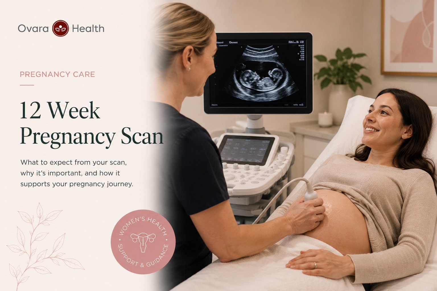
Wondering what to expect at a 12 week scan? The 12-week scan (also called the nuchal translucency scan) is one of the first detailed ultrasound examinations most pregnant women have during pregnancy.
Usually offered between 11weeks + 3 days and 13weeks +6 days of pregnancy, it confirms the pregnancy is progressing normally, establishes your estimated due date from a measurement called the crown-rump length and forms the first part of combined screening for chromosomal conditions including Down’s syndrome.
The scan takes around 20 to 30 minutes and is quite commonly performed through the abdomen in a painless procedure.
Key Takeaways
- The 12-week scan (nuchal translucency) is primarily a dating scan: crown-rump length gives your most accurate estimated due date
- Combined first trimester screening (scan plus blood test) detects around 90% of Down’s syndrome pregnancies
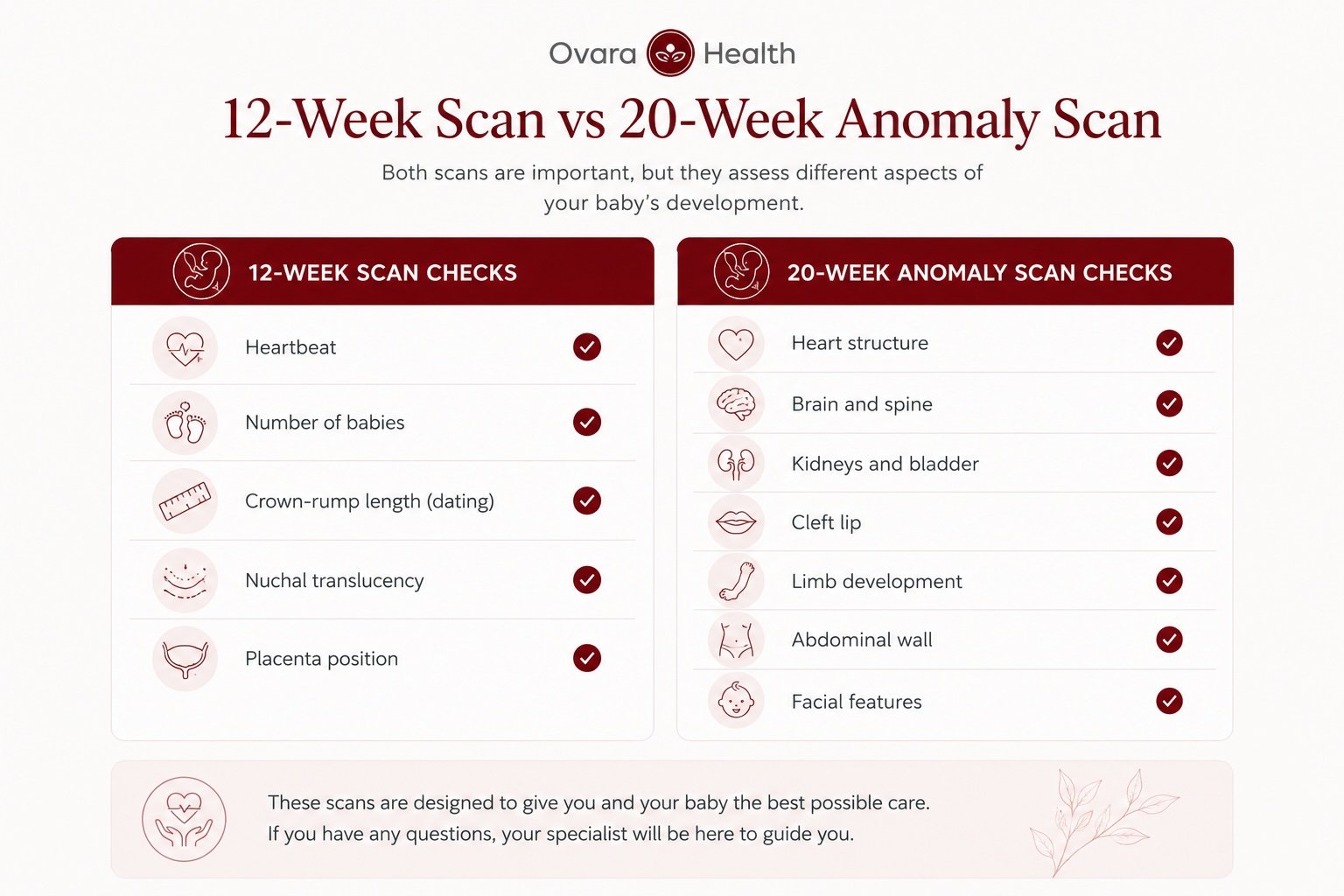
- The scan checks heartbeat, number of babies, placenta position and nuchal translucency (it is not a full anatomical survey)
- Most structural checks happen at the 20-week anomaly scan, not at 12 weeks
- Private 12-week scans are available at Ovara Health in London with same-week appointments and a consultant-reviewed report
What happens at the 12-week scan (nuchal translucency scan)?
You will lie on a couch with your abdomen exposed from your waistband to just below the ribs and the sonographer / Consultant applies a cold gel and moves a probe across your skin – the probe is firm but not painful.
Most women find the appointment entirely manageable, and many find it one of the more reassuring parts of early pregnancy.
How long does it take?
A standard NHS 12-week scan takes around 20 minutes whereas a private appointment, particularly one that includes a consultant consultation alongside the scan, typically runs for 30 to 45 minutes and is more thorough. If a blood test for combined screening is also required at the same visit this takes a little extra time.
What measurements does the sonographer take?
The primary measurement is the crown-rump length (CRL) – this is the distance from the top of the baby’s head to the base of the spine and is the most reliable way to date a pregnancy in the first trimester, with a margin of error of around five to seven days.
Your estimated due date is set from this measurement and if there is a discrepancy between the scan date and the date calculated from your last menstrual period, the scan measurement takes precedence.
Alongside the CRL, the sonographer will:
- Confirm a heartbeat is present and regular
- Check whether you are carrying more than one baby
- Measure the nuchal translucency (NT), the fluid at the back of the baby’s neck
- Note the position of the placenta
- Assess the volume of amniotic fluid around the baby
- Check basic anatomy: the shape of the head, the abdominal wall, the limb buds
Will it be internal or external?
Most 12-week scans are performed abdominally. An internal (transvaginal) scan is occasionally needed if the baby’s position makes abdominal imaging difficult, or if the scan is done at the earlier end of the window.
Internal scanning is safe in pregnancy and if it is needed, the sonographer will explain why and ask for your consent.
Does it hurt?
No. Ultrasound uses sound waves, not radiation, and the scan itself is painless. Some women find the pressure on a full bladder a bit uncomfortable and most scanning units ask you to arrive with a full bladder because it helps position the uterus for better imaging at this stage.
What can the 12-week scan detect?
The 12-week scan is primarily a dating and viability scan with the addition of chromosomal screening.
A detailed anatomical survey is done at 20 weeks – women sometimes leave the 12-week scan feeling reassured that everything is structurally normal, when in reality many structural assessments cannot be completed until the second trimester.
Down’s syndrome and chromosomal conditions
The nuchal translucency (NT) measurement is what most people are thinking about when they ask what the 12-week scan can detect.
Increased fluid at the back of the baby’s neck is associated with a higher probability of chromosomal conditions including Down’s syndrome (trisomy 21), Edwards’ syndrome (trisomy 18) and Patau’s syndrome (trisomy 13).
NT measurement alone is a risk indicator – not a diagnosis. A measurement of less than 3.5mm is generally within the normal range and above 3.5mm, the probability of a chromosomal condition increases, but it is important to note that the majority of pregnancies with a raised NT are chromosomally normal. Around 1 in 20 pregnancies will have an NT above this threshold.
When the NT measurement is combined with a blood test measuring PAPP-A and free beta-hCG alongside maternal age, the combined first trimester screening test detects approximately 90% of Down’s syndrome pregnancies, according to NHS screening guidance. That is significantly more accurate than the NT measurement used in isolation.
Structural markers visible at 12 weeks
Some structural problems can be identified at 12 weeks even though the detailed anatomy scan comes later. These include:
- Anencephaly (absence of the skull and normal brain development): reliably detectable at this stage
- Abdominal wall defects including gastroschisis and exomphalos, where abdominal organs develop outside the body
- Severe or absent limb development
- Large cystic structures such as a cystic hygroma at the back of the neck
These are uncommon findings. The large majority of 12-week scans show normal appearances across all of these markers.
What the 12-week scan cannot reliably tell you
A common misconception is that a normal 12-week scan means the baby is structurally healthy. In practice, many structural conditions are simply not visible at this gestation – the heart is too small for a detailed assessment at 12 weeks and the kidneys, spine and face are properly examined at 20 weeks. Cleft lip is not reliably detected at 12 weeks.
The question I hear most from patients at this stage is whether the scan can tell the sex of the baby and sometimes it can, but not reliably because at 12 weeks, the external genitalia are present but not sufficiently differentiated in most cases to give a confident answer.
Any estimate made at 12 weeks carries a real error rate. If finding out the sex matters to you, wait until the 20-week scan or book a dedicated gender scan from around 16 weeks onwards.
What is the combined first trimester screening test?
The combined test is part of the NHS antenatal screening programme, offered to all pregnant women. It uses three pieces of information together:
- The nuchal translucency (NT) measurement from the scan
- A blood test measuring two hormones: PAPP-A and free beta-hCG
- Your age, because the background risk for chromosomal conditions increases with maternal age
These are fed into a risk calculation that produces a probability score for Down’s syndrome, Edwards’ syndrome and Patau’s syndrome.
Understanding your result
Results are given as a ratio, for example 1 in 1,200 or 1 in 45. On NHS screening, a result of 1 in 150 or above is classified as “higher chance.” Below 1 in 150 is “lower chance.”
Higher chance does not mean your baby has a chromosomal condition – the majority of women with a higher-chance result go on to have chromosomally normal babies. A higher-chance result means further testing is offered: either chorionic villus sampling (CVS) or amniocentesis, which are diagnostic rather than screening tests.
In my experience, the waiting period between having the combined screening blood test and receiving the result is one of the most anxious parts of early pregnancy. Results typically come back within 7 to 10 days. If you are opting for combined screening, plan for that window. I make a point of mentioning this before the scan so patients are not caught off guard by the wait.
Can I decline combined screening?
Yes. Combined screening is offered and some women decline because they would not act on a higher-chance result whereas others want all available information to make an informed decision. There is no right answer and a good antenatal team will support whatever you choose without pressure in either direction.
For women who want a higher level of certainty, non-invasive prenatal testing (NIPT) offers around 99% detection for Down’s syndrome with a very low false positive rate.
NIPT analyses cell-free fetal DNA in maternal blood and can be done from 10 weeks. It is not currently available on the NHS as a first-line test but is widely available privately.
How should I prepare for my 12-week scan?
It is important to arrive with a full bladder and you will usually be asked to drink around 500ml to 1 litre of water in the hour before your appointment and to avoid going to the toilet before the scan. A full bladder pushes the uterus upward, which improves image quality in the first trimester.
It is helpful to wear comfortable, loose clothing as you will need to lower your waistband and expose the lower half of your abdomen. Fasting is not needed for the combined screening blood test.
If you have maternity notes or any earlier scan reports, bring them to the appointment. If you are attending a private clinic for the first time, bring any relevant medical history including previous pregnancy records.
What if something is found that needs further investigation?
Most 12-week scans are normal. The sonographer completes the measurements, checks the markers described above and confirms that appearances are as expected.
If an unexpected finding is identified, the next steps depend on what it is. A slightly raised NT measurement on its own changes the risk calculation but does not alter pregnancy management at that point whereas a suspected major structural abnormality will prompt referral to a fetal medicine unit for a more detailed assessment.
Unexpected findings at 12 weeks, particularly borderline NT measurements are not rare and many do not result in a significant problem. In a private setting, if a serious concern is identified, referral to a fetal medicine specialist can typically be arranged within days.
NHS 12-week scan vs private: what is the difference?
The NHS dating scan is offered free of charge to all pregnant women in the UK and a standard appointment typically lasts around 20 minutes. Waiting times between referral and appointment can be two to four weeks depending on your area.
A private 12-week scan offers several things the standard NHS pathway does not:
- Same-week or next-day availability with flexible hours including evenings and weekends
- Longer appointments of 30 to 45 minutes
- Higher-resolution imaging in many cases
- A detailed written report at or shortly after the appointment
- Direct access to a consultant obstetrician or gynaecologist alongside or reviewing the scan
- Time for questions without time pressure
At Ovara Health in Chelsea, private pregnancy scans are available with direct access to our specialist Consultants without a GP referral.
Early pregnancy scans start from £200 and an initial consultation with a viability scan with the consultant is available from £450.
When to consider a private 12-week scan
A private scan is worth considering if:
- You want to be seen quickly rather than waiting several weeks for an NHS appointment
- You have had a previous pregnancy loss and want a consultant-level appointment
- You want a consultant-reviewed written report
- You are considering NIPT and want to discuss your options in the same appointment
- You want more time for questions than a standard NHS appointment allows
- Heavy vaginal bleeding with severe cramping
- Sudden, severe one-sided abdominal pain (a possible sign of ectopic pregnancy)
- Shoulder tip pain combined with abdominal pain and dizziness
- Fainting or collapse
Call your midwife, GP or early pregnancy unit the same day if you notice any vaginal bleeding in early pregnancy, a significant reduction in pregnancy symptoms or abdominal pain that does not settle.
Frequently asked questions
How many weeks pregnant should I be for the 12-week scan?
The 12-week scan is offered between 11 weeks and 13 weeks and 6 days of pregnancy. For combined first trimester screening, the scan needs to take place between 11 weeks and 13 weeks and 6 days.
Will I be able to see my baby clearly at 12 weeks?
Yes. By 12 weeks the baby is well-formed and clearly visible on ultrasound. You will see movement, the heartbeat and the outline of the head, body and limbs.
Can the 12-week scan detect Down’s syndrome?
The 12-week scan, when combined with a blood test as part of combined first trimester screening, detects approximately 90% of Down’s syndrome pregnancies. The scan alone is not diagnostic. Definitive diagnosis requires CVS or amniocentesis.
What abnormalities can be seen at the 12-week scan?
The 12-week scan can identify certain major structural problems including anencephaly, abdominal wall defects and severe limb abnormalities – it is not a full structural survey. The detailed anatomy scan at 20 weeks assesses a much wider range of features.
Is the 12-week scan the same as the dating scan?
Yes. The terms refer to the same appointment.
Does the 12-week scan use radiation?
No. Diagnostic ultrasound uses high-frequency sound waves. There is no ionising radiation involved.
Can I find out the sex of my baby at the 12-week scan?
Not reliably. A dedicated gender scan from around 16 weeks is more accurate.
What happens if the nuchal translucency measurement is higher than normal?
An NT above 3.5mm increases the risk calculation for chromosomal conditions. Further testing will be discussed. Most pregnancies with a raised NT are chromosomally normal.
Do I need a GP referral for a private 12-week scan?
No. At Ovara Health, you can book directly without a referral.
How long does the 12-week scan take?
An NHS scan takes around 20 minutes. A private appointment with a consultation takes 30 to 45 minutes.
What should I do if I cannot attend my NHS 12-week scan?
Contact your midwife or antenatal department as soon as possible. The combined screening window closes at 13 weeks and 6 days.
Is the 12-week scan linked to the 20-week anomaly scan?
Yes. Both are part of the NHS antenatal screening programme. The 12-week scan focuses on dating and chromosomal screening; the 20-week scan is the detailed structural survey.
Sources
- NHS: Ultrasound scans in pregnancy
- Tommy’s: 12-week ultrasound scan information
- RCOG: Ultrasound scans in pregnancy
- NICE Guideline NG201: Antenatal care
This article is for general information only and does not constitute medical advice. It is not a substitute for assessment by a qualified clinician.
Book a Private 12-Week Pregnancy Scan in London
At Ovara Health in Chelsea, you can book a private 12-week scan with direct access to our pregnancy & gynaecology Consultants. Same-week appointments are available, no GP referral is needed and a written consultant-reviewed report is included.
Book your 12-week scan at Ovara Health
Early pregnancy scans from £200. Consultation with viability scan from £450. Call us on 0207 751 4488.